The Nobel Prize in
Chemistry 2014
Sulekha Rani.R ,PGT Chemistry,KV NTPC kayamkulam
The Nobel Prize in Chemistry 2014 was awarded
jointly to
Eric Betzig, Stefan
W. Hell and William E. Moerner
"for the development of super-resolved
fluorescence microscopy".
Two US and one German scientist win Nobels for opening a window into the nanoworld with their development of ‘super-resolved fluorescence microscopy’
Here’s one of the winners, Eric Betzig, speaking in Singapore in 2012:
“I’d like to give my talk on a personal level, so you can see at least how science is done by a guy like myself who doesn’t consider himself a scientist at all.”
Surpassing the limitations of the light microscope
For a long time optical microscopy was held back by a presumed
limitation: that it would never obtain a better resolution than half the
wavelength of light. Helped by fluorescent molecules the Nobel Laureates in
Chemistry 2014 ingeniously circumvented this limitation. Their ground-breaking
work has brought optical microscopy into the nanodimension.
In
what has become known as nanoscopy, scientists visualize the pathways of
individual molecules inside living cells. They can see how molecules create
synapses between nerve cells in the brain; they can track proteins involved in
Parkinson’s, Alzheimer’s and Huntington’s diseases as they aggregate; they
follow individual proteins in fertilized eggs as these divide into embryos.
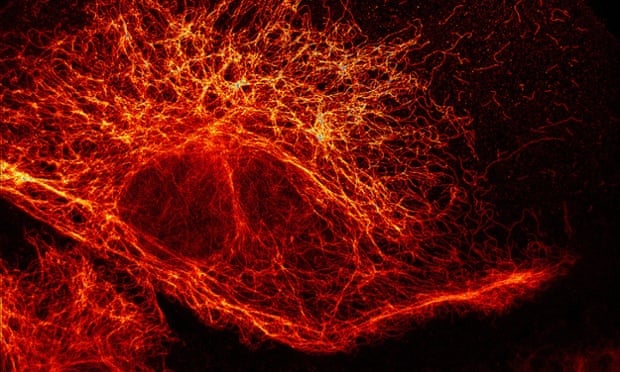
A network of filaments in a mammalian cell revealed by fluorescence microscopy. Photograph: Stefan W Hell/Division of Optical Nanoscopy/German Cancer Research Center
It
was all but obvious that scientists should ever be able to study living cells
in the tiniest molecular detail. In 1873, the microscopist Ernst Abbe
stipulated a physical limit for the maximum resolution of traditional optical
microscopy: it could never become better than 0.2 micrometres. Eric Betzig, Stefan W. Helland William E. Moerner are awarded
the Nobel Prize in Chemistry 2014 for having bypassed this limit. Due to their
achievements the optical microscope can now peer into the nanoworld.
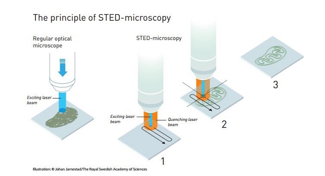
Two
separate principles are rewarded. One enables the method stimulated
emission depletion (STED) microscopy, developed by Stefan Hell in 2000. Two
laser beams are utilized; one stimulates fluorescent molecules to glow, another
cancels out all fluorescence except for that in a nanometre-sized volume.
Scanning over the sample, nanometre for nanometre, yields an image with a
resolution better than Abbe’s stipulated limit.
Eric
Betzig and William Moerner, working separately, laid the foundation for the
second method, single-molecule microscopy. The method relies upon
the possibility to turn the fluorescence of individual molecules on and off.
Scientists image the same area multiple times, letting just a few interspersed
molecules glow each time. Superimposing these images yields a dense super-image
resolved at the nanolevel. In 2006 Eric Betzig utilized this method for the
first time.


In 1873, Ernst Abbe discovered that there was a seemingly insurmountable limit to how powerful an optical microscope can be. To put it very simply, he found that light can’t get around the bends on tiny objects. According to his calculations, the wavelengths of visible light meant that the maximum limit of the resolution of an optical microscope is 0.2 micrometers, which is about half a wavelength. For over a century, this blocked the lower limit for optical studies, which prevented the direct observation of structures on a nanoscale, such as individual molecules.


What the new Laureates did wasn’t a violation of Abbe’s limit, but more of a workaround. The basic idea was that if light couldn’t be made to bend around nano-sized objects, then the nano-objects could be made to radiate their own light. It’s like the difference between trying to spot a tiny object by shining a giant searchlight on a field, and leaving the field dark but decorating the object with tiny Christmas lights. This insight led to the development of two new methods of what is now called nanomicroscopy.
Today,
nanoscopy is used world-wide and new knowledge of greatest benefit to mankind
is produced on a daily basis.
MORE ABOUT THE METHODS
1. STED Microscopy
The first method is called STimulated Emission Depletion (STED) microscopy, which was discovered by Stefan Hell in 2000, though he’d been working on the problem since his student days in the 1990s. The key to the method was to avoid the diffraction limit by not just using molecules that fluoresce, but to stimulate them to do so using a laser.
Hell’s technique used fluorescent molecules, such as antibodies that link to specific structures; for example, DNA strands. When these are hit with a pulse of light, they glow in return. Normally, this produces an image like a cluster of glowing wool, but the STED microscope solves this problem by using a pair of lasers. One sets the molecules glowing, and the other eliminates all but the desired molecules from the picture.
As the first laser scans over a specimen and sets the molecules aglow, a second laser follows close behind. This is tuned so that larger molecules absorb the laser light, causing them to discharge their energy and cease glowing. The nano-sized molecules are too small to be affected and keep giving off light. The result is an image of extremely fine detail.
2. Single Molecule Microscopy
The second method is called Single-Molecule Microscopy. It was developed Eric Betzig and William Moerner while working independent of one another, and was used by Betzig for the first time in 2006. It's based on the ability to see a single fluorescent molecule and using this to build up an image of extremely fine detail.
In 1989, W. E. Moerner was the first to measure the light absorption of a single molecule. This was followed in 1997, when Moerner and Roger Tsien at the University of California in San Diego were studying green fluorescent proteins (GFP). Moerner discovered that it was possible to turn this fluorescence of GFPs on and off by using different wavelengths of light. By shining light with a wavelength of 488 nanometers, he could get it to glow, and this would die and not return when shone on again. However, by hitting the molecule with light at 405 nanometers, the molecule would revive and glow again.
This basic idea was expanded upon by Moerner and Betzig independently, to create a new microscopy technique that exploited the on/off system. In this, a specimen would be treated with a number of different fluorescent molecules that bonded to different molecular structures, but always at least 0.2 micrometers apart. Each of these would glow at different times as stimulated. By activating each fluorescent molecule type in turn, then switching them off, a series of different images were produced
These images were then scanned individually and subjected to a statistical algorithm that sharpened each one. When the images were combined in layers, the result was a single image showing complex, high-resolution structures.
Aside from removing the need for a lot of squinting, these methods have removed the theoretical lower limit to optical microscopy. According to the Nobel Foundation, they are already finding applications, such as the ability to look at individual molecules instead of studying "average" molecules in bulks numbering millions. They are currently being used to study synapses in Alzheimer’s and Huntington’s disease, and to gain a better understanding of protein development in embryos.